FIGURE 6:
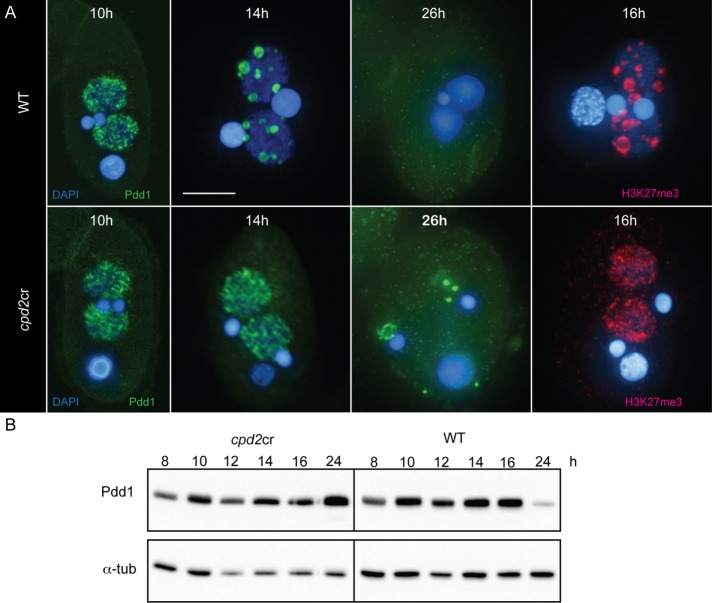
cpd2cr cells do not form elimination bodies. (A) In WT cells, Pdd1-bound, heterochromatin-marked IESs coalesce into large foci at 14–16 h after induction of mating, as indicated by immunofluorescence staining with anti-Pdd1 or anti-H3K27me3 antibodies. In cpd2cr cells, these foci do not form and Pdd1 persists as abnormal cellular aggregates. Scale bar, 5 μm. (B) Western blotting also shows abnormal persistence of Pdd1 expression. Protein extracts were made from aliquots of mating cpd2cr or WT cells processed at the indicated time points after induction of mating.

