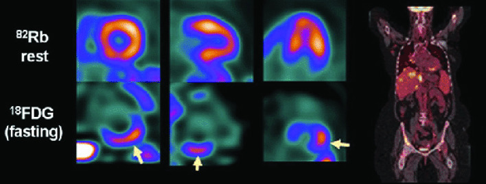
Figure 1.
PET/CT images in a patients with cardiac sarcoidosis. Perfusion imaging (above, arrow) show hypoperfusion in the basal inferior wall, corresponding with increased FDG uptake (under, arrow). Fused whole-body coronal imaging shows the systemic extent of sarcoidosis. Reprinted with permission from Springer, from Caobelli F and Bengel FM. J Nucl Cardiol 2015;22: 971–974. FDG, fluorodeoxyglucose; PET, positron emission tomography.

