Figure 2.
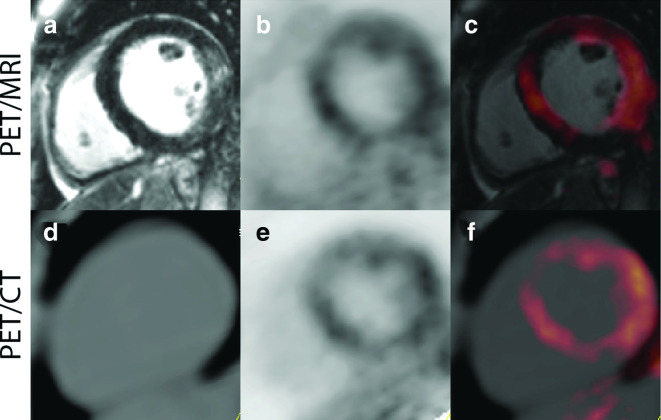
Representative Images from a Patient with cardiac sarcoidosis, imaged with PET/MR (A–C) and PET/CT (D–F). There is evidence of enhanced signal seen on the delayed enhancement MR images in the lateral wall (A), and the increased 18F-FDG uptake in the septal, anterior, and lateral regions on both the PET/CT and PET/MR images (C, F). This patient had an ejection fraction of 49% with mild global hypokinesis, without wall motion abnormalities. Reprinted under the terms of the Creative Commons Attribution 4.0 International License (http://creat ivecommons.org/licenses/by/4.0/) from Wisenberg G et al. J Nucl Cardiol. 2019. doi: 10.1007/s12350-018-01578-8 [56]. No changes were made. 18F-FDG, 18F-fluorodeoxyglucose; PET, positron emission tomography.

