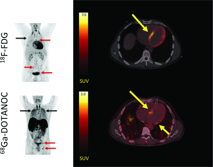
Figure 4.
Representative 18F-FDG PET/CT and 68Ga-DOTANOC PET/CT images in a patient with cardiac sarcoidosis. Left panel: MIPs showing dilated cardiomyopathy and multiple 18F-FDG and 68Ga-DOTANOC avid lymph nodes (red arrows). In addition, there is massive and diffusely increased activity in the lung parenchyma (black arrows) representing active pulmonary sarcoidosis. Right panel: while 18F-FDG PET/CT was inconclusive due to failed suppression of tracer uptake from the myocardium (top), 68Ga-DOTANOC images showed a clearly pathological uptake in the septum (bottom). Reprinted under the terms of the Creative Commons Attribution 4.0 International License (http://creat ivecommons.org/licenses/by/4.0/) from EJNMMI Res. 2016;6(1):52 [82]. No changes were made.18F-FDG, 18F-fluorodeoxyglucose; PET, positron emission tomography.

