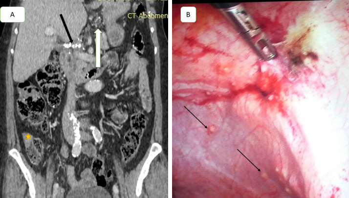
Figure 3.

A 57-year-old female with primary mucinous ovarian cancer. (A) Coronal contrast-enhanced CT scan obtained during venous phase demonstrates multiple small soft tissue peritoneal nodules at region 2 (lesser omentum nodules white arrow), calcified soft tissue deposit at liver hilum (black arrow), and soft tissue serosal implant on ascending colon (star), CT PCI = 11. Laparoscopic image (B) showed multiple small soft tissue nodules scattered in lateral peritoneal fold (missed by CT), laparoscopy and pathology PCI = 9, CT falsely upstaged the case from low to moderate group, patient underwent optimal cytoreduction. PCI, peritoneal cancer index.
