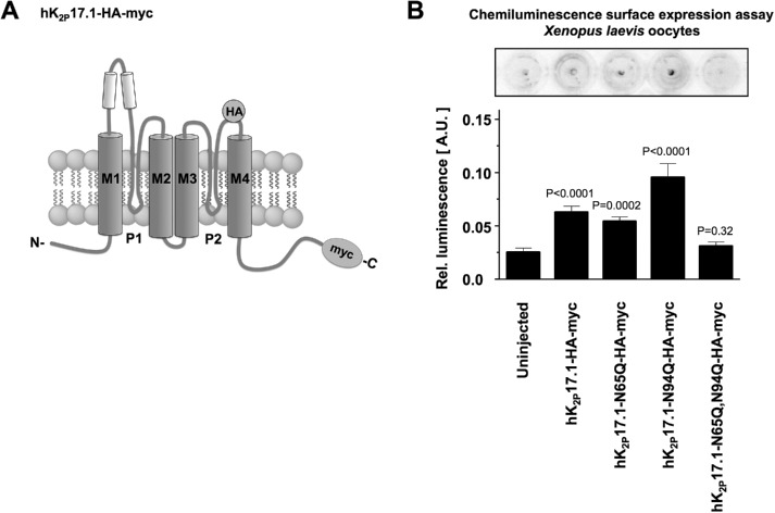
FIGURE 4:
N-glycosylation regulates surface expression of hK2P17.1. (A) Schematic diagram of the hK2P17.1-myc-HA construct used in this experiment. An internal HA tag localized at the extracellular part of the P2-M4 interdomain was used for immunological detection of hK2P17.1 dimers at the surface of nonpermeabilized Xenopus oocytes. (B) Surface expression of WT hK2P17.1 and mutants was measured by HRP-mediated chemiluminescence in Xenopus oocytes. Data are given as mean values ± SEM of n = 11– 29 cells, p values are indicated above the bars.

