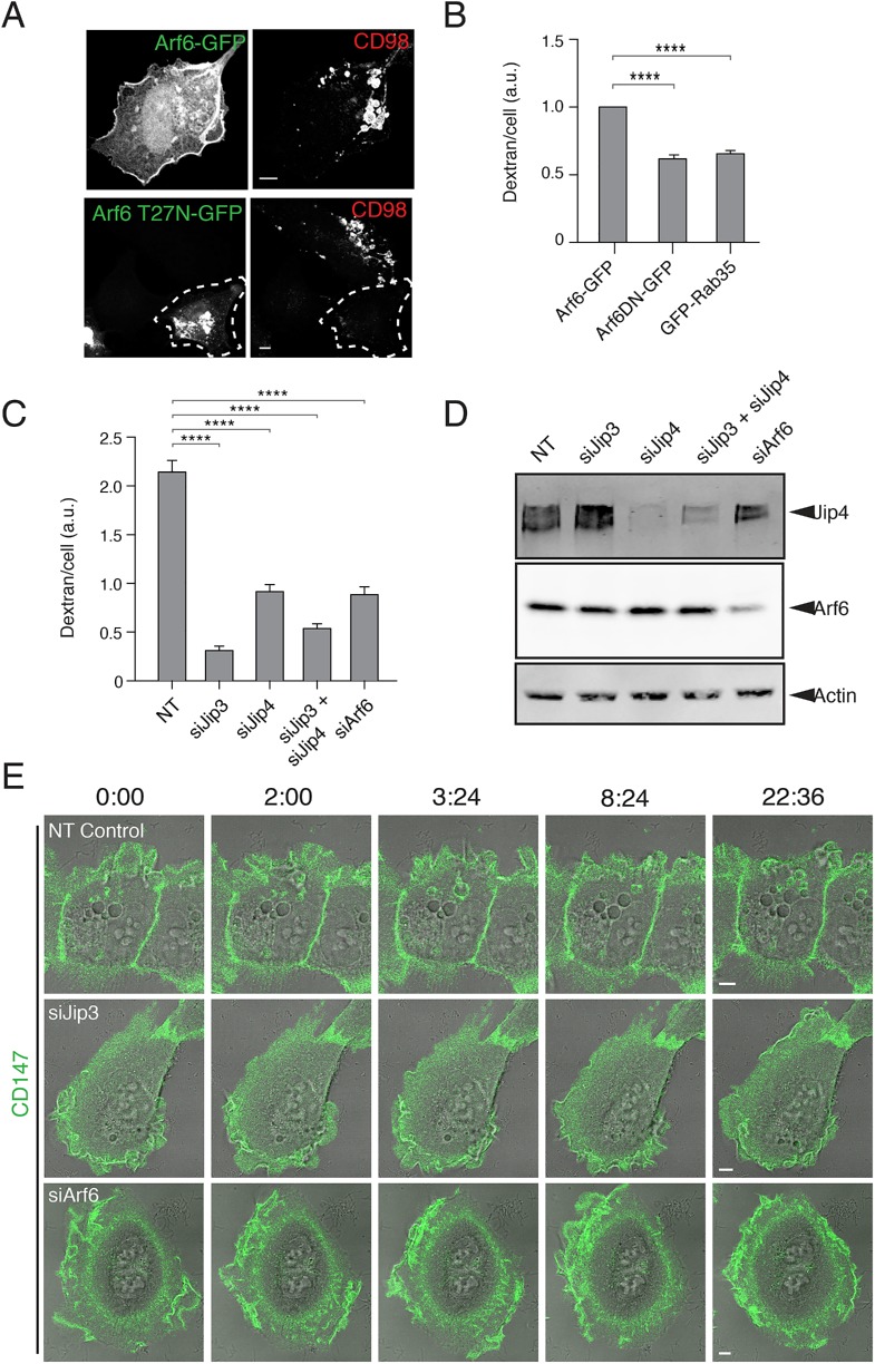
FIGURE 5:
Arf6 and JIP3/JIP4 are required for macropinosome formation and maturation. (A) HT1080 cells expressing Arf6-GFP (top) or Arf6T27N-GFP (bottom) were incubated with antibodies to CD98 for 20 min prior to acid wash, fixation, and immunolabeling with fluorophore–conjugated secondary antibodies. (B) HT1080s cells expressing GFP, Arf6T27N-GFP, or GFP-Rab35 were incubated with Dextran-647 for 20 min, and dextran uptake was measured by flow cytometry. Bar graphs show the geometric mean dextran fluorescence of GFP-positive cells, reported as a fraction of Arf6-GFP control, from three separate experiments (GFP-positive cells were gated from a total of 100,000 cells per experiment). An ordinary one-way ANOVA was used to compare groups to the Arf6-GFP control. Error bars represent ± SD. ****p value < 0.0001. Bars, 5 μm. (C) Control HT1080 cells and cells depleted of JIP3, JIP4, JIP3, and JIP4, or Arf6 were incubated for 20 min with dextran-594 and then fixed and imaged. Bar graphs show the average dextran fluorescence from 240 individual cells per condition in a single matched experiment, error bars show the standard error of the mean. Experiments were repeated three times with similar phenotypes. An ordinary one-way ANOVA was used to compare groups to the NT control. ****p value < 0.0001. (D) The extent of siRNA knockdown of JIP3, JIP4, JIP3 and JIP4, and Arf6 was assessed by Western blot of JIP4 and Arf6. (E) HT1080 cells depleted by siRNA of nontargeting control (Supplemental Movie 5), JIP3 (Supplemental Movie 6), or Arf6 (Supplemental Movie 7) were incubated with Alexa488–conjugated primary antibodies against CD147 for 1 h prior to live-cell imaging. Membrane ruffling and macropinocytosis were followed in live-cell imaging over the course of 20 min.

