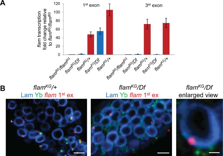
FIGURE 2:
flam first exon transcripts are retained and form perinuclear foci in flamKG/Df ovaries. (A) RT-qPCR analysis of flam transcription in flamBG and flamKG mutant ovaries using primer pairs for the first or third exons. Fold change of flam transcription in the ovaries with indicated genotypes relative to flamBG/flamBG ovaries, normalized on the Adh transcript levels, is indicated along the Y-axis (mean ± SD for two replicates). The flam third exon transcripts were barely detected in flamBG and flamKG mutants, but the flam first exon transcripts are retained in flamKG mutant. (B) RNA FISH (red) with flam first exon probe coupled with immunostaining of Yb bodies with anti-Yb antibody (green) and nuclear envelope with anti-lamin Dm0 antibody (blue) in the follicle cells of the stage 2 egg chamber of flamKG/+ (left panel), flamKG/Df (central panel), and enlarged view of flamKG/Df (right panel). Scale bars are 5 μm in the left and central panels and 0.7 μm in the right panel.

