FIGURE 1:

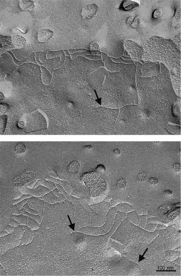
Freeze fracture electron microscopic images of MDCK II cells reveals basal free ends and strand breaks; the apical surface showing microvillar stubs is at the top of the images. Some free ends are indicated by arrows. Bar 100 nm.

Freeze fracture electron microscopic images of MDCK II cells reveals basal free ends and strand breaks; the apical surface showing microvillar stubs is at the top of the images. Some free ends are indicated by arrows. Bar 100 nm.