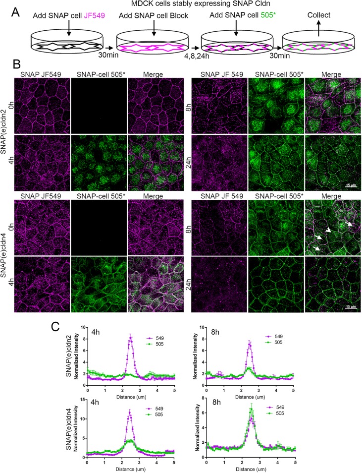
FIGURE 4:
Pulse-chase SNAP tag labeling of SNAP(e)cldn2 and 4 reveals that newly synthesized cldns are trafficked to the lateral membranes and that cldn2 is biosynthesized and/or trafficked more slowly than cldn4. (A) Scheme of SNAP tag labeling, first with JF549 SNAP-ligand to label all cldn2 or 4 (T = 0), followed by blocking for 30 min with SNAP-cell block. This is followed by incubation for various periods of time (4, 8, and 24 h) and then labeling newly synthesized cldns with SNAP-cell 505*. (B) Fluorescence of SNAP ligand labeling of SNAP(e)cldn2 (top panels) and SNAP(e)cldn4 (bottom panel) expressing MDCK II cells. Cells are imaged after labeling with JF549 SNAP ligand and then labeled and imaged with 505*at 4, 8, and 24 h after blocking. Difference in biosynthesis/trafficking is most evident at the 4-h time point. Arrows indicate vesicular colocalization of old and new SNAP(e)cldn4; arrowhead indicates vesicular structure containing only new SNAP(e)cldn4. (C) Line scan across cell contacts at 4 and 8 h reveal more accumulation of new SNAP(e)cldn4 than SNAP(e)cldn2; normalization was performed as described in Materials and Methods; n = 14 line scans.

