
Figure 4.
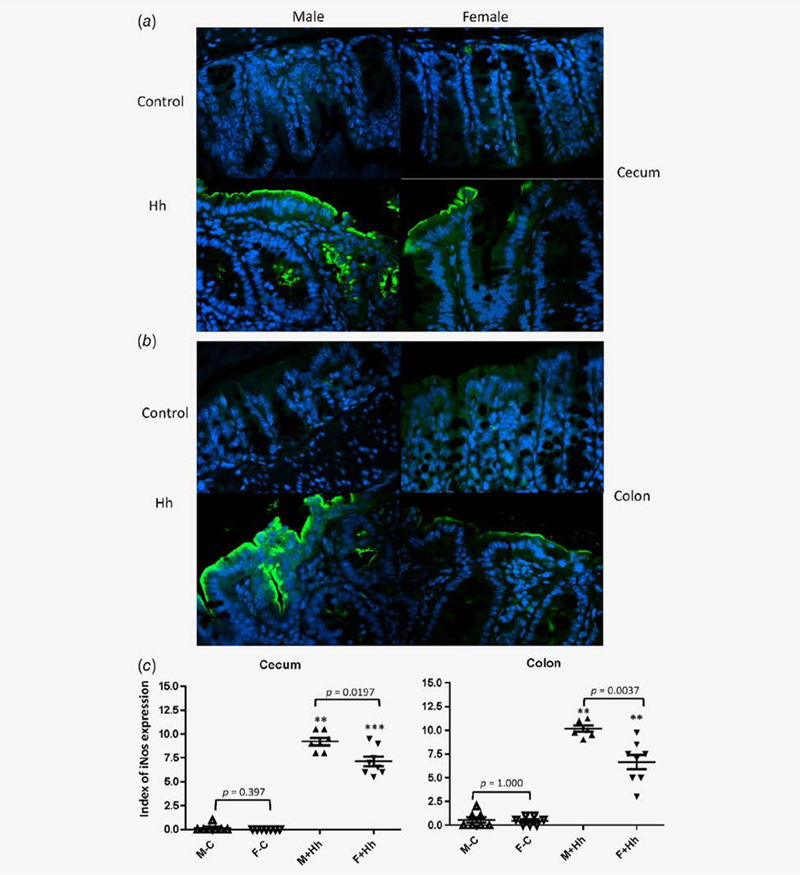
Epithelial iNos expression was significantly elevated in the lower bowel of H. hepaticus-infected RagIl10gpt males vs. H. hepaticus- infected RagIl10gpt females. The representative iNos staining images: the ceca (a) and colons (b). (c) Index of iNos epithelial cells in the lower bowel of RagIl10gpt mice. H. hepaticus infection significantly elevated iNos expression compared to the uninfected controls. *represents p values when compared to the sham controls: **<0.01, ***<0.001.