
Figure 6.
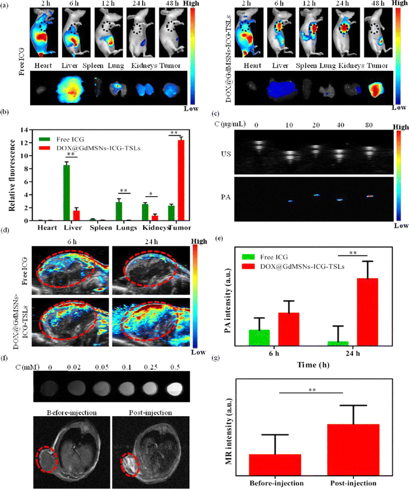
Multimodal applications of FIGS: a In vivo NIR fluorescence images after intravenous injection of free ICG and DOX@GdMSNs-ICG-TSLs in tumor-bearing mice at 2, 6, 12, 24, and 48 h, and the corresponding fluorescence images of different tissues after treated with free ICG and DOX@GdMSNs-ICG-TSLs at 24 h. b Relative fluorescence intensity of ICG in major organs induced by 808 nm laser (1.5 W/cm2) irradiation at 24 h after i.v. administration. c US and PA images of DOX@GdMSNs-ICG-TSLs at various concentrations of ICG. d PA images of tumor-bearing mice after 6 and 24 h intravenous injection via tail of free ICG and DOX@GdMSNs-ICG-TSLs, respectively. e PA intensity of tumor sites after treatment with free ICG and DOX@GdMSNs-ICG-TSLs at 6 and 24 h. f T1-weighted MR images (7 T, spin–echo sequence; repetition time, TR = 500 ms; echo time, TE = 14.92 ms) of DOX@GdMSNs-ICG-TSLs nanoparticles at various Gd concentrations. And T1-weighted MR images of tumor-beating mice before and after injected with DOX@GdMSNs-ICG-TSLs for 24 h. g Relative MR intensity before and after injecting DOX@GdMSNs-ICG-TSLs. Data are presented as means ± SD (n = 5); *P < 0.05, **P < 0.01. (Adapted with permission from Sun Q, You Q, Wang J, et al (2018) Theranostic Nanoplatform: Triple-Modal Imaging-Guided Synergistic Cancer Therapy Based on Liposome-ConjugatedMesoporous Silica Nanoparticles. ACS Appl Mater Interfaces © 2018 American Chemical Society).