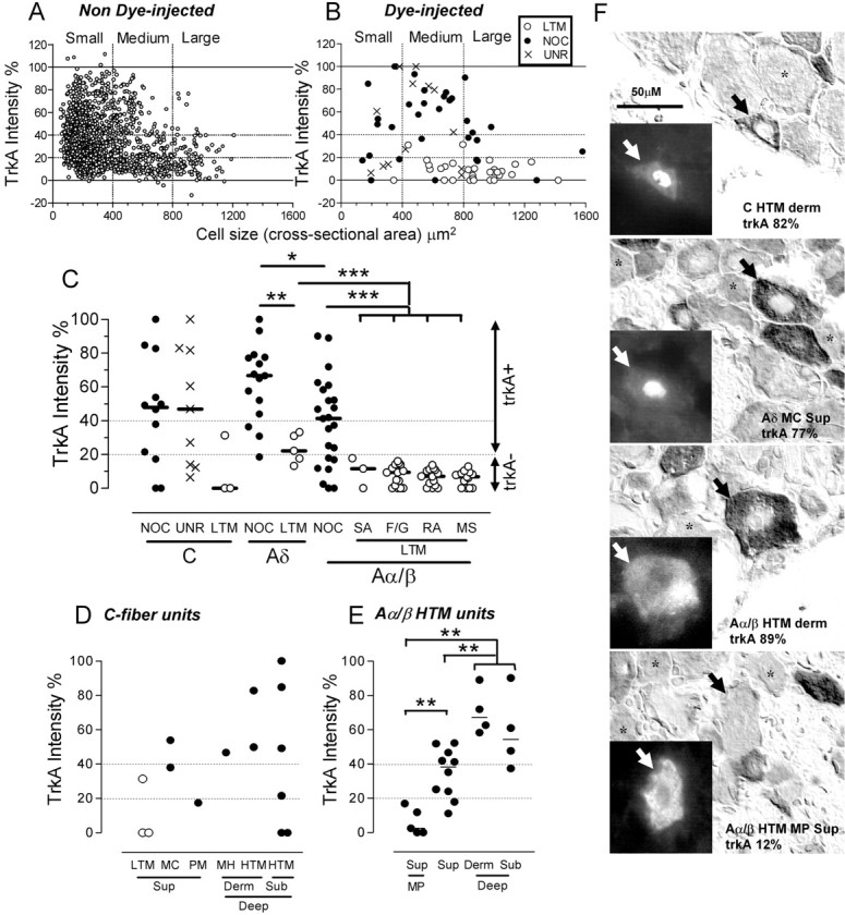
Figure 1.

A, B, Sizes of all non-dye-injected neuronal profiles with nuclei in sections of one L5 DRG from each of three rats (A) compared with those of identified dye-injected neurons (B). Vertical dotted lines indicate boundaries between small, medium, and large neurons. Horizontal dotted lines indicate borderlines between negative and positive neurons and between weakly positive (20-40%) and strongly positive (≥40%) neurons. C, trkA intensities of physiologically identified DRG neurons; a Kruskal-Wallis test comparing medians of the three groups of nociceptors shows a significant difference between Aδ and Aα/β neurons and no differences between the four groups of Aα/β LTMs; Mann-Whitney U tests show significant differences between Aδ nociceptors and Aδ LTM neurons, between Aα/β nociceptors and LTMs, and between Aδ and all Aα/β LTMs. CLTM neurons are excluded, because n = 3. D, trkA intensity in CLTMs and populations of C nociceptive neurons. E, Comparisons of trkA intensities of Aα/β-fiber HTM subgroups. Moderate pressure (MP) units had significantly lower trkA intensities than other HTMs with superficial or deeper (dermal and subcutaneous together) receptive fields, and HTMs with superficial receptive fields had significantly lower intensities than units with deep receptive fields (Mann-Whitney tests). F, Photomicrographs show fluorescent images of representative dye-injected neurons in insets to left: dyes were Lucifer yellow, cascade blue, and for the two lowest cells ethidium bromide; note the typically nuclear fluorescence with cascade blue and Lucifer yellow. The same neurons stained for trkA, under interference contrast optics, are indicated by arrows (right). Asterisks indicate examples of trkA- neurons. The top three injected neurons are strongly trkA+, and the last is a trkA-MP unit. The scale bar (50 μm; top left image) applies to all photomicrographs, including insets. For C and E, *p < 0.05, **p < 0.01, ***p < 0.001. NOC, Nociceptive; UNR, C-fiber unresponsive; SA, slowly adapting; F/G, field or guard hair; RA, rapidly adapting; MS, muscle spindle; MC, mechano-cold; PM, polymodal; MH, mechano-heat. Depth in tissues: Sup, superficial; Derm, dermal; Sub, subcutaneous.
