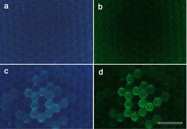
Figure 7.
Fluorescence microscopic photographs of the eyes of female (a, b) and male (c, d) P. rapae crucivora, at the level of the corneal facet lenses, under UV (a, c) and violet (b, d) excitation. Whereas the fluorescence in female eyes is more or less homogeneous and faint, that of type II ommatidia in male eyes is distinct [for the relationship of the fluorescence and the ommatidial types, see Qiu et al. (2002)]. Scale bar, 50 μm.

