
Figure 1.
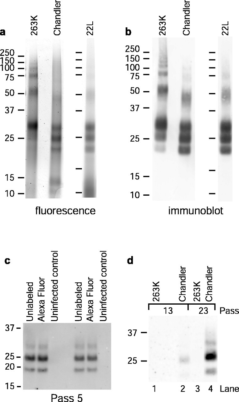
Fluorescence labeling of PrP-res and chronic infection of SN56 cells. a, SDS-PAGE and fluorescence detection of PK-treated PrP-resA568 preparations. b, Immunoblots of the samples shown in a using anti-PrP antibody D13. c, Comparison of long-term PrP-res production in cells infected with A568-labeled or unlabeled PrP-res. Cells at ∼30-40% confluency in a 96-well plate were exposed to 20 ng of PrP-res or PrP-resA568 in 30 μl of medium (0.67 ng/μl PrP-res), incubated for 5 h, fed with an additional 150 μl of medium, incubated for 2 d, and passed into a T-24 well for another 4 d of growth (pass 1). Cells were passed at a 1:10 dilution from this point. At pass 5, PrP-res was precipitated from cell lysates and assayed by immunoblot with D13. Each sample was derived from lysate aliquots containing 224 μg of total protein. d, Long-term PrP-res production in cells treated with the designated types of PrP-resA568 under conditions identical to those used in the confocal microscopy experiments described below and in Materials and Methods. Compared with the protocol used in c, the concentration of PrP-resA568 (0.032 ng/μl) was much lower, and the time of exposure before the change of medium (4 h) was much shorter, resulting in lower overall levels of PrP-res in the cultures. A total of 500 μg of total lysate protein equivalents was loaded per lane. The results in c and d show that A568-labeled PrP-res remained infectious for SN56 cells.