
Figure 1.
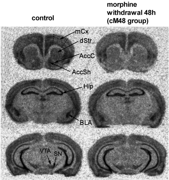
In situ hybridization autoradiograms showing α-synuclein mRNA signal in the brains of control mice and mice subjected to chronic morphine treatment followed by 48 h of withdrawal. AccC, Nucleus accumbens core; AccSh, nucleus accumbens shell; dStr, dorsal striatum; Hip, hippocampus; mCx, motor cortex; SN, substantia nigra. Note the reduction in the signal densities in the dStr, AccC, AccSh, BLA, and cortex.