Figure 2.
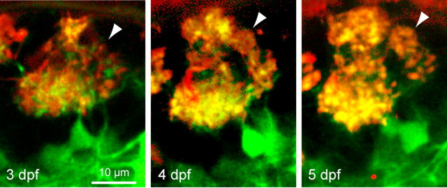
Time-lapse imaging of a protoglomerulus. Two-photon optical section through a protoglomerular structure in the same individual at three subsequent developmental days. Red, DiA labeling; green, dlx4/6::GFP expression. The arrowheads depict a small glomerular structure that appears to segregate from a preexisting protoglomerulus.

