
Figure 4.
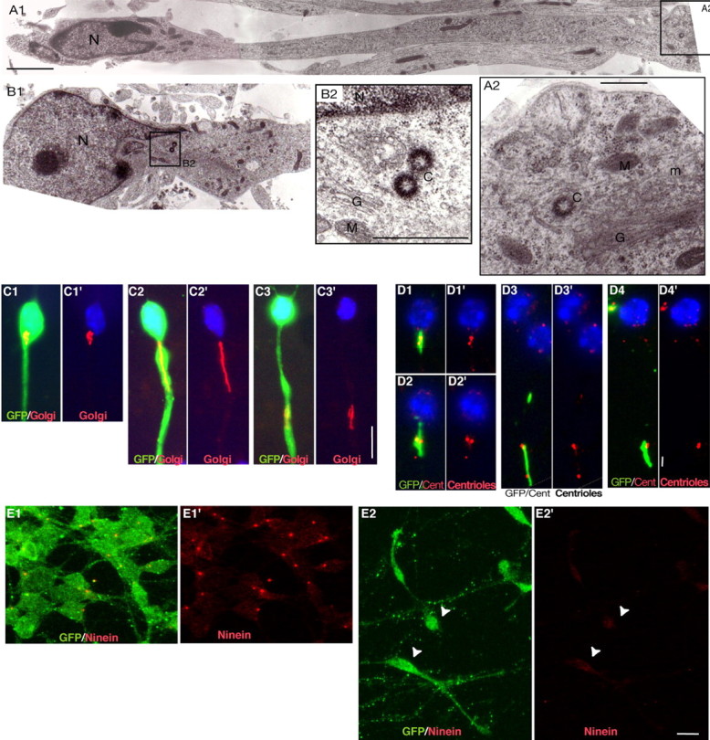
Localization and morphology of the centrosome and Golgi apparatus in MGE cells migrating on cortical cells. A1-B2, Ultrastructural analysis of MGE cells migrating on cortical axons reveals the presence of the centriole(s) and Golgi apparatus in a swelling of the leading process that can be located distant from (15 μm; A1) or close to (B1) the nucleus. The nucleus (N) occupies one pole of the cell and shows an indentation oriented toward the leading process (Gregory et al., 1988). Swellings contain one or two centrioles (C), depending on the plane of section (A2, B2). Centrioles were always located near the Golgi apparatus (G). M, Mitochondria; ER, rough endoplasmic reticulum; m, microtubules. Scale bars: A1, 2 μm; A2, 500 nm; B2, 200 nm. C1-C3′, Immunostainings of GFP (green; C1-C3) and the medial compartment of the Golgi apparatus (CTR433 in red) in migrating MGE cells. Nuclei are labeled with bis-benzimide (blue). The Golgi apparatus is located either close to the nucleus (C1′) or rostrally in a swelling of the neuritic trunk (C2′, C3′). It shows a compact (C1′, C3′) or a linear (C2′) conformation. Scale bar, 10 μm. D1-D4′, Immunostainings of the Golgi apparatus (CTR433 in green; D1-D4) and centrioles [centrin-2 (Cent) in red]. The two centrioles are associated with the Golgi apparatus (GA). In cells with a compact GA, the two centrioles appear as two close dots (D1′, D2′) or a single larger dot (D4′). In cells with a linear GA, the two centrioles are widely separated and line up along the GA (D3′). Scale bar, 2 μm. E1-E2′, In GFP-positive MGE cells aggregated on laminin (E1, E1′; GFP in green), ninein antibodies (red) strongly label a single dot in the centrosome. In MGE cells migrating on cortical axons (E2, E2′; GFP in green), the ninein-positive dot is no longer visible, whereas a faint labeling of the whole soma can be observed (E2′; white arrowheads). Scale bar, 10 μm.