Figure 6.
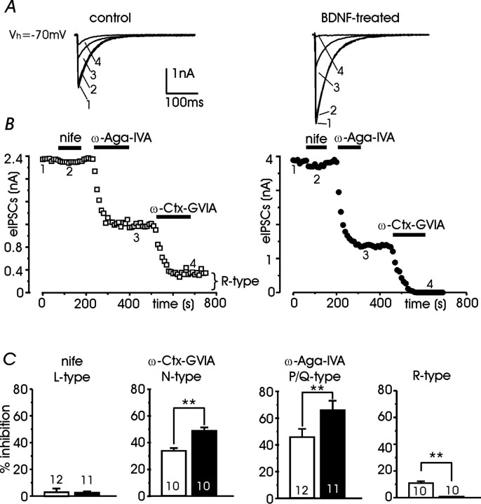
Pharmacological dissection of presynaptic Ca2+ channel types supporting eIPSCs. A, Examples of eIPSCs recorded before (1) during sequential application of 3 μm nifedipine (nife) (2), 0.5 μm ω-Aga-IVA (3), and 1 μm ω-Ctx-GVIA (4) in control neurons (left) and treated neurons (right). B, Time course of eIPSC amplitude relative to the same neurons shown in A. C, Mean percentage contribution of Ca2+ channel types to eIPSCs estimated by separately applying the three Ca2+ antagonists on control neurons (open bars) and treated neurons (filled bars); **p < 0.01 vs controls.

