
Figure 1.
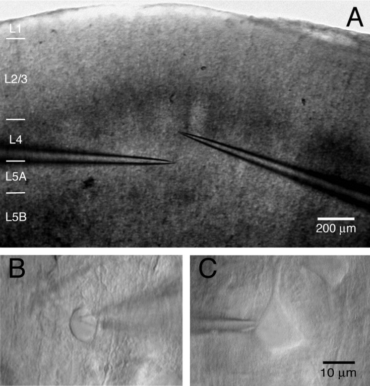
IR-DIC image of an L4-to-L5A connection. A, IR-DIC image of a coronal slice with clearly discernible barrels. The pipettes mark the position of the presynaptic cell (in layer 4) and the postsynaptic cell (in layer 5A). Note that both the presynaptic and the postsynaptic neurons are located at the border of the barrel column. B, High-magnification image of the presynaptic spiny stellate cell. C, Postsynaptic L5A pyramidal cell. Scale bar in C also applies to B.