
Figure 2.
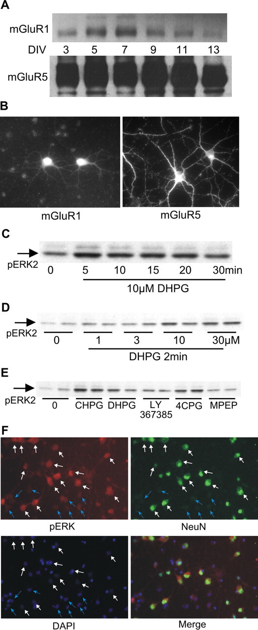
Group I metabotropic glutamate receptors stimulate ERK2 phosphorylation in E18 striatal neurons. A, Western blot analysis of mGluR1 and mGluR5 expression in cultured striatal neurons as a function of DIV. Protein content per lane for these blots was identical. Film exposure times were also identical to demonstrate the magnitude of the difference in protein levels of mGluR1 versus mGluR5. B, Immunofluorescence using subtype-specific antibodies shows that both mGluR1 and mGluR5 are localized in somata and processes, with mGluR5 more abundant than mGluR1. C, Time course of ERK2 phosphorylation by DHPG. D, Concentration-response of ERK2 phosphorylation by DHPG. E, Pharmacological characterization indicates that mGluR5 is the predominant group I mGluR mediating ERK2 phosphorylation in striatal neurons. mGluR agonist DHPG (10 μm) or CHPG (750 μm) was applied for 2 min. Receptor antagonist LY367385, 4CPG, or MPEP (see concentrations in Results) was applied 30 min before stimulation with DHPG. F, Cells were stimulated with DHPG and double labeled using anti-phosphoERK with anti-NeuN to mark neurons. White arrows indicate cells positive for NeuN; blue arrows indicate cells negative for NeuN. DAPI staining indicates the location of all cells.