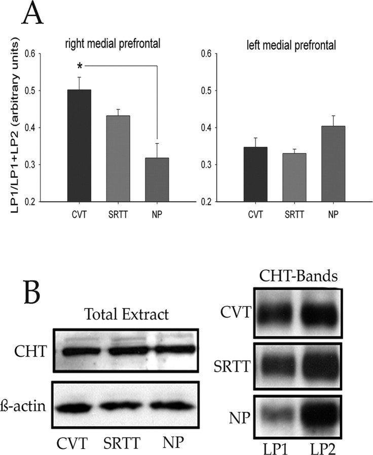
Figure 3.
A, An increased proportion of CHTs was found in the plasma membrane-enriched fraction obtained from the right mPFC of CVT-performing rats when compared with NP rats (the asterisk depicts the significant difference indicated by post hoc multiple comparisons using Tukey's honestly significant difference). Data from the left mPFC, left or right posterior cortex, or striatum did not differ between groups. B, Left bands, Total CHT protein levels were not affected by performance in either task. β-Actin levels were monitored to verify the amount of samples used for immunoblot analysis. Right bands, CHT activity in LP1 and LP2 fractions, indicating the greater proportion of CHTs in the plasma membranes in the right mPFC of CVT-performing rats when compared with NP rats.

