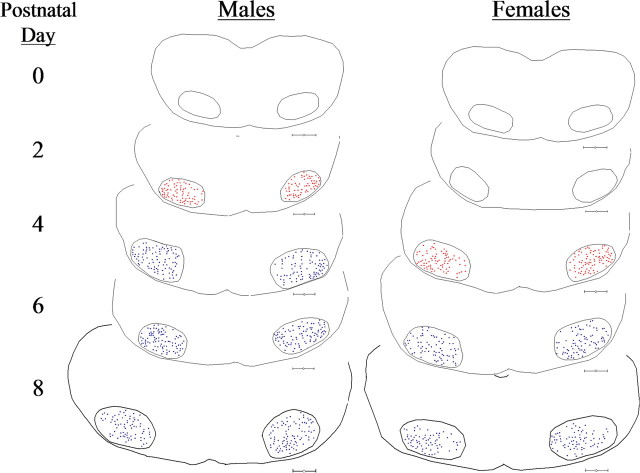
Figure 5.
AR expression first occurs in male hamster FMNs by P2 and is delayed by 2 d in females. Neurolucida tracings of brainstem sections containing bilateral facial nuclei from an AR immunocytochemistry developmental time-course study in P0, P2, P4, P6, and P8 male and female hamsters are shown. Red dots represent cytoplasmic-only FMN labeling. Blue dots represent cytoplasmic plus nuclear FMN labeling. Scale bars, 300 μm.

