Figure 5.
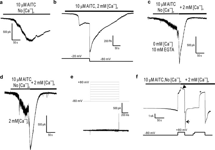
Calcium-induced potentiation and inactivation of TRPA1 is voltage dependent, and the inactivation is reversed by sustained depolarization. a, b, In the absence (a) of external calcium, AITC elicits only slow activation (-80 mV holding potential), whereas in the presence (b) of external calcium, inactivation is complete at hyperpolarized (-80 mV) holding potentials but not at depolarized (-20 mV) potentials. c, Addition of calcium to the external medium of slowly activated TRPA1 channels elicits fast potentiation followed by inactivation (-80 mV holding potential). Notice that intracellular calcium is being chelated with 10 mm EGTA. d, The calcium-induced potentiation and subsequent inactivation of TRPA1 channels also occurs with high (2 and 3 mm) levels of internal calcium (introduced by diffusion from the pipette, under the whole-cell configuration, over a period of >5 min). e, Whole-cell recordings of TRPA1-expressing HEK cells exposed to several voltage steps reveal no voltage-dependent gating of TRPA1 channels in the range of -80 to +80 mV. f, However, calcium-induced inactivation, which is reduced at positive (+80 mV) membrane potentials (arrowhead) and maximal at negative (-80 mV) ones (arrow), is reversed by positive holding potentials (asterisk) after a delay of ∼30 s. The mechanism for this recovery from inactivation is presently unknown. No [Ca2+]o solutions contain 10 mm EGTA as chelator and have no CaCl2 added. For all panels, the trace shown is a representative of at least four separate cells.

