
Figure 7.
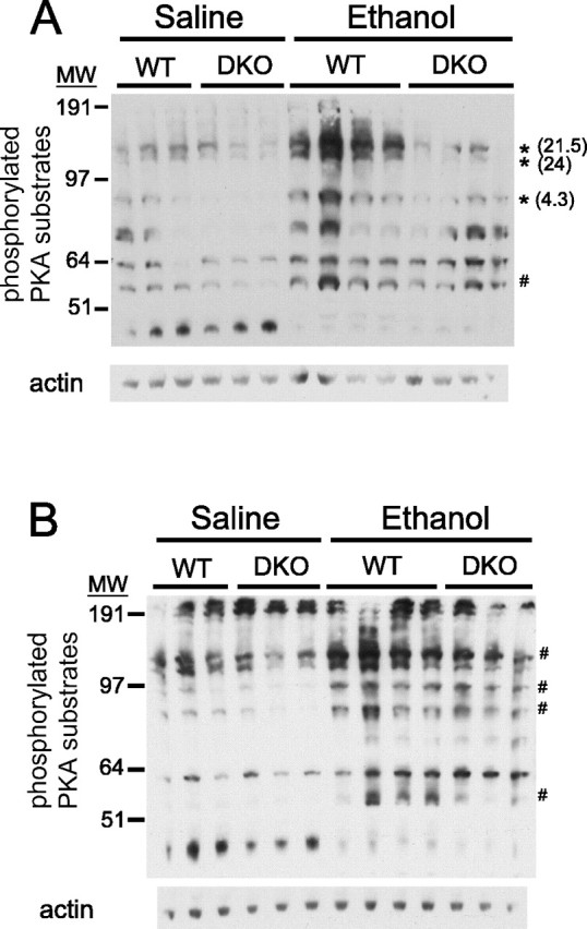
Ethanol-induced phosphorylation of a subset of PKA substrates is compromised in the cortex, but not the cerebellum, of DKO mice. Western blots of lysates of cortex (A) or cerebellum (B) isolated from WT and DKO mice treated with ethanol or saline are shown, indicating increased intensity of several bands detected by an antibody to the phosphorylated PKA consensus motif after ethanol treatment. The asterisk indicates bands that were increased after ethanol treatment to a greater extent in the WT brain than in the DKO brain (treatment, p < 0.05; genotype, p < 0.05). The number in parentheses indicates the fold difference in mean intensity of indicated bands in WT ethanol group versus DKO ethanol group. The # symbol indicates bands that were increased equivalently in WT and DKO brain after ethanol treatment (treatment, p < 0.05; genotype, p > 0.05). There were no bands in cerebellar samples that were increased after ethanol treatment to a greater extent in WT mice than in DKO mice. MW, Molecular weight.