
Figure 3.
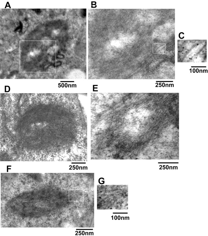
Immunoelectron microscopy analysis of inclusions in transfected cells. Cells were cotransfected with myc-synphilin-1 and α-synuclein, S129A α-synuclein, or S129E α-synuclein, and 72 h later, cells were subjected to immunoelectron microscopy. A and D-F are representative images of different shape cytoplasmic inclusions in cells coexpressing myc-synphilin-1 and wild-type α-synuclein. B represents a higher magnification of the rectangle in A. C and G represent the higher magnification of rectangles in B and F, which shows the gold-labeled filaments by using phospho-α-synuclein antibody.