Figure 8.
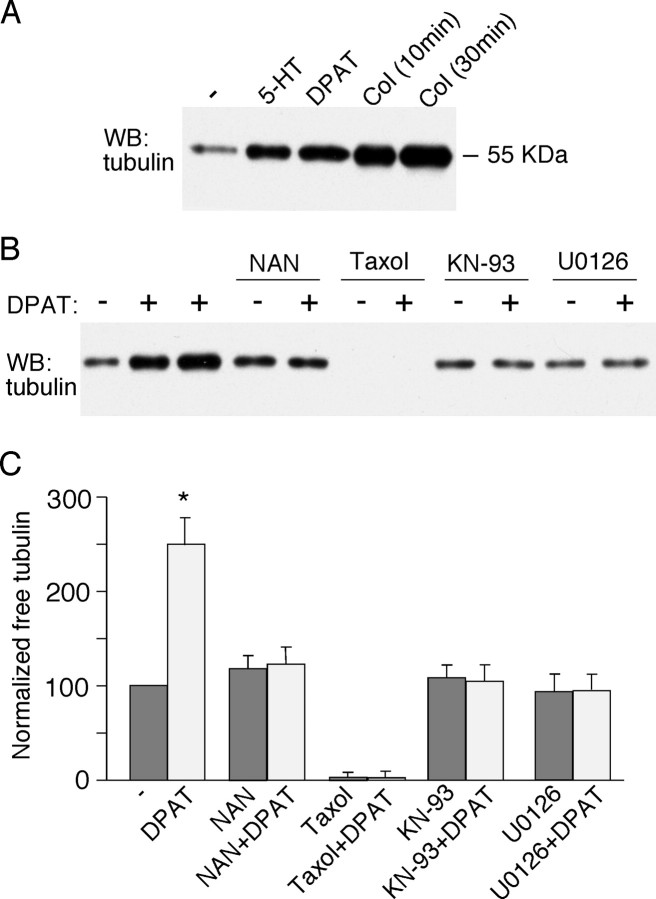
Activation of 5-HT1A receptors induces an increase in free tubulin. A, Western blot (WB) analysis of free tubulin in lysates of cultured PFC neurons treated without (-) or with (+) 5-HT (40 μm, 10 min), 8-OH-DPAT (DPAT; 40 μm, 30 min), or colchicine (Col; 30 μm, 10 or 30 min). B, Western blot analysis of free tubulin in lysates of cultured PFC neurons treated without (-) or with (+) 8-OH-DPAT (40 μm, 30 min; lane 2, 5 min treatment) in the absence or presence of various agents (added 10 min before 8-OH-DPAT treatment), including the 5-HT1A receptor antagonist NAN-190 (NAN; 40 μm), the microtubule stabilizer taxol (10 μm), the CaMKII inhibitor KN-93 (10 μm), and the MEK inhibitor U0126 (20 μm). C, Quantification of free tubulin assay. Free tubulin level was normalized to control (-), based on the intensity of the free tubulin band from Western blot analyses. Each point represents mean ± SEM of four to five independent experiments. *p < 0.001, ANOVA.

