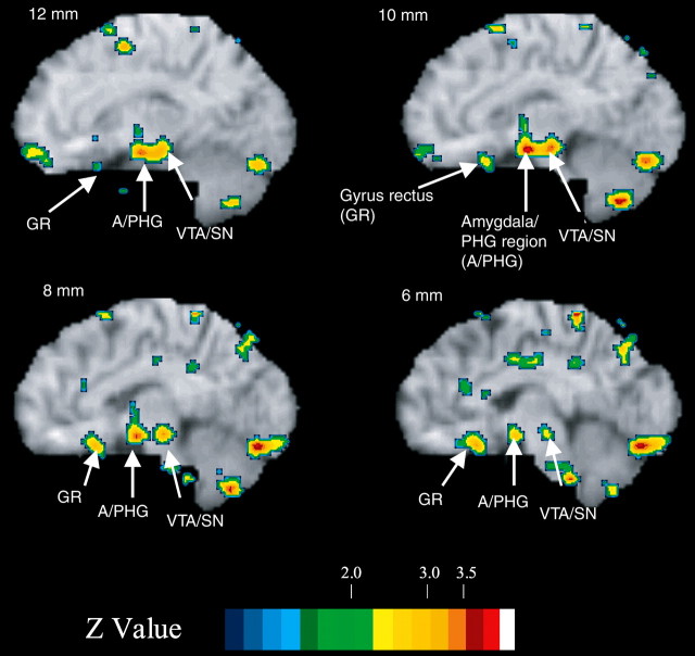
Figure 2.
Sagittal slices for neural activations in the contrast between TEST and CONTROL under interoceptive feedback during training. Changes in activity are overlaid on sagittal slices of a standard brain MRI. Color scales show activations (+) and deactivations (-) in Z scores. Slices are shown in millimeters. Note that TEST requires inference based on review of the feedback history without feedback stimulation; feedback stimulation occurred only during training. PHG, Parahippocampal gyrus.

