
Figure 6.
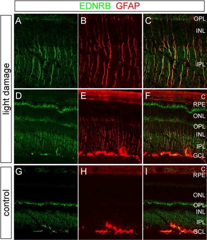
Accumulation of EDNRB and GFAP in Muller cells in light-damaged retinas as determined by immunostaining. A-F, BALB/c mice were exposed to 6 h of bright light with pupil dilation, followed by a 24 h recovery in darkness. A-C, Enlarged views of the central retina from D-F. G-I, Control BALB/c mice maintained in darkness. Light exposure leads to the accumulation of EDNRB and GFAP in Muller cells, characterized in the central retina by their radial fibers. EDNRB also accumulates in presumptive astrocytes in the GCL and at the outer tips of the Muller cells, which abut the photoreceptor inner segments between the outer nuclear layer and the RPE. Among Muller cells in the light-damaged retina, both EDNRB and GFAP show substantial cell-to-cell variability. C, Choroid; ONL, outer nuclear layer; OPL, outer plexiform layer; IPL, inner plexiform layer.