
Figure 1.
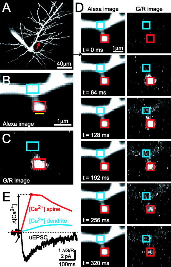
Simultaneous two-photon glutamate uncaging and two-photon [Ca2+] imaging in single dendritic spines. A, CA1 pyramidal neuron filled with Alexa 594. B, Blow-up of a dendritic branch (arrow in A). The yellow bar indicates the position of the uncaging beam. Rectangles denote ROIs for analysis of Δ[Ca2+] in the spine head (red) and parent dendrite (blue). C, G/R image of the [Ca2+] response to glutamate uncaging, averaged over two poststimulus frames (same spine as in B). D, One [Ca2+] imaging trial in frame scan mode (dark frame and the first baseline frame are not shown). The arrow indicates the timing of the uncaging stimulus (same spine as in B, C). E, Uncaging-evoked [Ca2+] transient (ΔG/R) in the spine head (red) and parent dendrite (blue). The simultaneously recorded NMDA-R uEPSC (black) is also shown. The spine is the same as in B-D. Each trace is an average of five trials.