Figure 4.
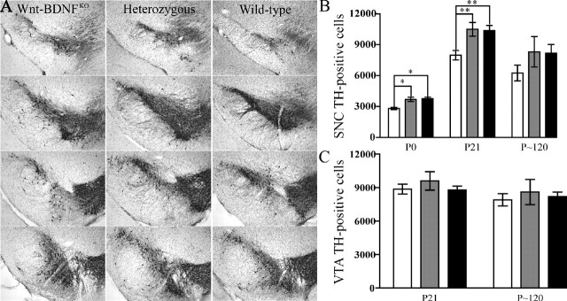
Wnt-BDNFKO mice have reduced TH expression in the SNC but not in the VTA. A, The extent of the SNC appears reduced in P21Wnt-BDNFKO mice compared with controls, and the number of TH fibers also seems to be reduced. The most anterior section is at the top, and the most posterior section is at the bottom. Coronal cryostat sections (40 μm) taken at 240 μm intervals stained for TH and counterstained with cresyl violet are shown. B, Optical fractionator estimates of the number of DA neurons in both hemispheres of the SNC of Wnt-BDNFKO mice and controls at P0, P21, and P120 (P0, n = 3/genotype; P21, n = 9, 8, and 9 for Wnt-BDNFKO, heterozygous, and wild type, respectively; P120, n = 3/genotype; *p < 0.05; **p < 0.01; one-way ANOVA with a Newman-Keuls posthoc test). C, There is no significant reduction in the total number of TH-positive cells of both hemispheres of the VTA of Wnt-BDNFKO mice compared with controls (P21, n = 11, 7, and 8 for Wnt-BDNFKO, heterozygous, and wild type, respectively, p = 0.6454; P120, n = 3/genotype, p = 0.8170). White bars, Wnt-BDNFKO; gray bars, heterozygous; black bars, wild type.

