
Figure 2.
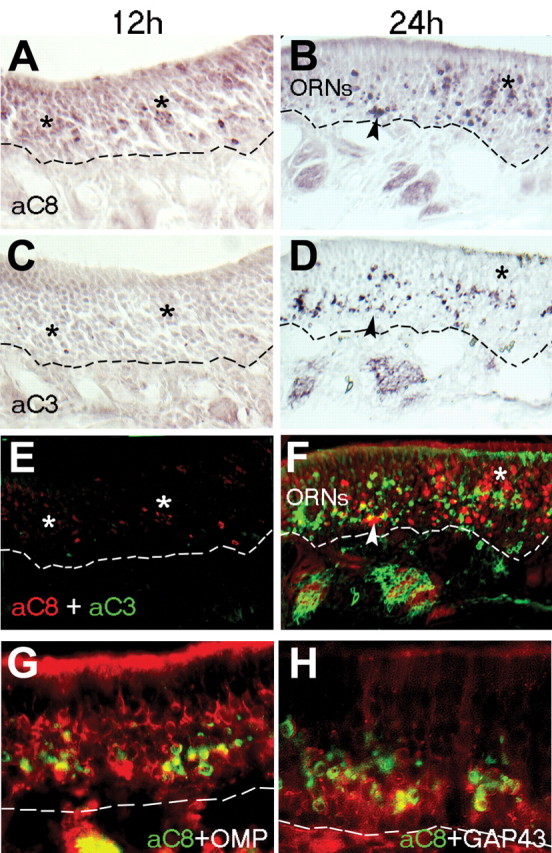
Caspase-8 activation peaks ahead of, and is more widespread than, active caspase-3 after bulbectomy. A, C, E, At 12 h postbulbectomy (12 h), active caspase-8 (A; aC8) is detected in a greater proportion of cells (asterisks) in the OE than active caspase-3 (aC3) on an adjacent section (C), a pattern emphasized when both images are overlaid (E; red, active caspase-8; green, active caspase-3). B, D, F, At 24 h postbulbectomy (24 h), some ORNs shared active caspase-8 (B; F, red) and caspase-3 (D; F, green) in common (arrowheads), but a greater proportion of ORNs in the more mature, apical layers of OE contain active caspase-8 alone (asterisks). G, H, In immunolabeled coronal sections of OE at 24 h postbulbectomy, active caspase-8 (green) is detected in both OMP-positive mature ORNs (G, red) and immature neurons expressing GAP43 (H, red). ORNs, Mature neuronal layers of olfactory epithelium. The dashed line indicates basal lamina.