
Figure 3.
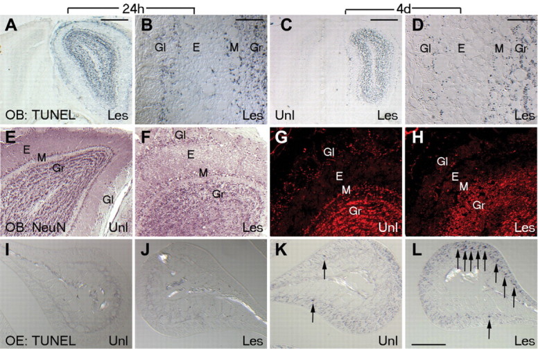
Olfactory bulb and epithelial neurons undergo apoptosis after NMDA lesion. A-D, Coronal sections of unilaterally NMDA-lesioned olfactory bulb show TUNEL-positive apoptotic cells restricted to the lesioned side only at 24 h (A, B) and 4 d (C, D) after NMDA infusion. TUNEL-positive neurons in several neuronal layers of the olfactory bulb (periglomerular, mitral, and granule) can be seen on the lesioned side but are relatively absent from the unlesioned side. E-H, Coronal sections of unilaterally NMDA-lesioned bulb at 24 h (E, F) and 4 d (G, H) immunolabeled for NeuN show disruption of neuronal layering in olfactory bulb at 24 h and disappearance of many bulb cells, notably in the mitral layer by 4 d. I-L, TUNEL labeling in the OE (shown on endoturbinate Iib; sample area 2 in Fig. 1 B) at 24 h and 4 d after NMDA lesion. Lesion-induced TUNEL-positive ORNs seen at 4 d (K, L) are not seen in the unlesioned side or in sham animals. E, External plexiform; Gl, glomerular; Gr, granule layers; Les, lesioned side; M, mitral/tufted; Unl, unlesioned side.