
Figure 6.
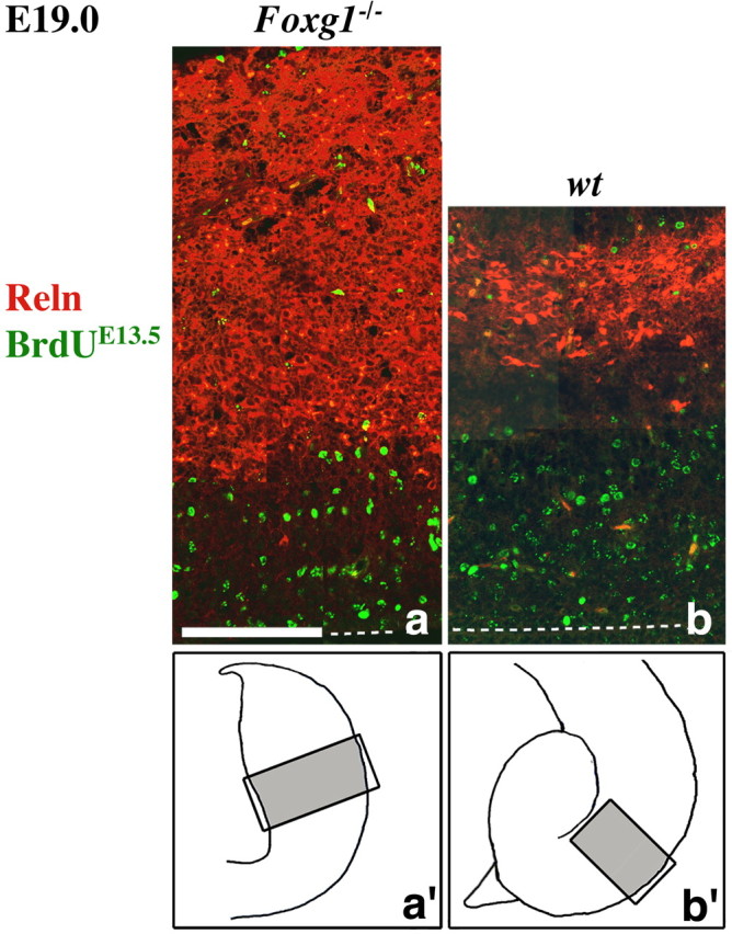
Neuronal birthdating in the Foxg1-/- cortical primordium. The distribution of BrdU (green) and Reln (red) on frontal telencephalic sectors of E19.0 Foxg1-/- (a) and wild-type (b) embryos, pulsed by BrdU at E13.5 is shown; ventricular is to the bottom and marginal is to the top. In a and b, the dashed lines indicate the ventricular border of the cortical wall. In a′ and b′, the silhouettes of the two telencephalons are shown, with boxes demarcating the sectors represented in a and b, respectively. Scale bar, 100 μm.