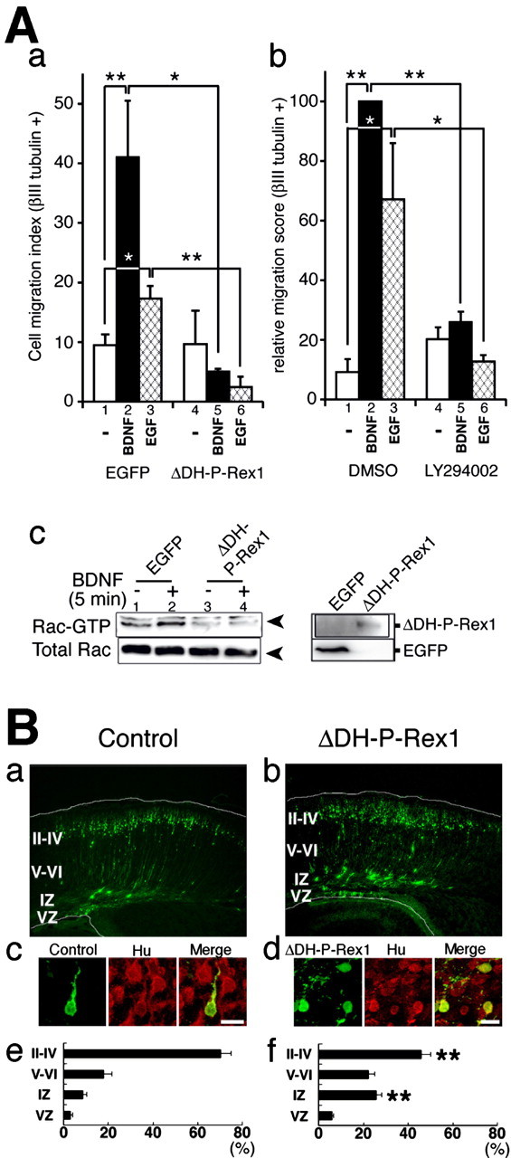
Figure 8.

A potential role of P-Rex1 in the migrating neurons. A, P-Rex1 is involved in BDNF- and EGF-induced trans-well migration of primary cortical neurons. a, pEGFP or pcCAG-EGFP-ΔDH-P-Rex1 plasmid was transfected into primary cultured cells derived from E14 mouse cerebral cortices. After incubation at 37°C for 16 h, trans-well migration assays were performed for 4-5 h with or without 10 ng/ml BDNF or 100 ng/ml EGF stimuli. Cells that migrated out on the bottom side of the filter were stained with the neuron-specific anti-βIII-tubulin antibody, and positive-cells were counted. Cell migration indices were calculated as the compensated cell number estimated at 30% transfection efficiency. b, A PI3-kinase inhibitor blocked BDNF- and EGF-induced trans-well migration of primary cortical neurons. Primary cultured cells derived from E14 mouse cerebral cortices were applied to each trans-well migration chamber. Trans-well migration to 10 ng/ml BDNF or 100 ng/ml EGF was assayed with or without 50 μm LY294002. Relative migration score was calculated as the relative percentages of the compensated cell number to that of the standard experiment (Ab, lane 2). All assays were repeated at least three times. Data were evaluated by using Student's t test. *p < 0.05; **p < 0.01. c, GTP-bound Rac1 in primary cultured cells transfected with an EGFP or ΔDH-P-Rex1 with or without administration of BDNF. pEGFP (lanes 1, 2) or pcCAG-EGFP-ΔDH-P-Rex1 (lanes 3, 4) was transfected into dissociated cells derived from E14 mouse cerebral cortices. After transfection, cultured cells were maintained in EBSS for 16 h and treated with or without BDNF for 5 min. The top left panel indicates GTP-bound Rac1. The bottom left panel shows total Rac1. The right box depicts EGFP and EGFP-ΔDH-P-Rex1 proteins. B, Effects of ΔDH-P-Rex1 on development of the cerebral cortex. E14 embryonic brains were transfected with a control vector (a, c, e) or a ΔDH-P-Rex1-expressing vector (b, d, f) by in utero electroporation, together with pEGFP to visualize the transfected cells. a, b, At P0, frozen brain sections were examined for EGFP fluorescence. White lines represent pial and ventricular surfaces. By immunostaining with an anti-tag antibody, expression of ΔDH-P-Rex1 was confirmed in GFP-positive cells (data not shown). c, d, Expression of a neuronal marker, Hu. E14 embryos were electroporated and killed at P0. Frozen sections were immunostained with anti-GFP (green) and anti-Hu (red) antibodies (c in the cortical plate; d in the intermediate zone). Scale bars, 10 μm. e, f, Estimation of cell migration by recording fluorescence intensities of EGFP in distinct parts of the cerebral cortex (Kawauchi et al., 2003). Each bar represents the mean ± SE percentage of relative intensities; n = 11. Asterisks indicate significant differences between the scores of the intermediate zone or layers II-IV of the cortical plate in control and ΔDH-P-Rex1-transfected brains (p < 0.01; t test). II-IV, Layers II-IV of the cortical plate; V-VI, layers V-VI of the cortical plate; IZ, intermediate zone; VZ, ventricular zone.
