
Figure 3.
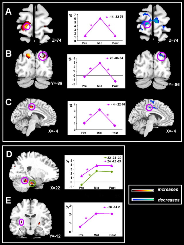
Anatomical localizations of coordination effort-related increases in activation across learning on a representative normalized brain. Temporary increased activation was observed in the left primary motor cortex/dorsal premotor area (A), right superior occipital area (B), and left posterior cingulate sulcus (C). Sustained increases compared with the PRE level were found in the right cerebellar lobule III and V (D) and left putamen/globus pallidus (E). Each graph represents the estimated signal changes for the most significant voxel in each area for the Bim-R contrast across all three scan sessions. An asterisk indicates a significant change for Bim > UniL + UniR at p < 0.05 after correction for multiple comparisons, with k > 10.