
Figure 3.
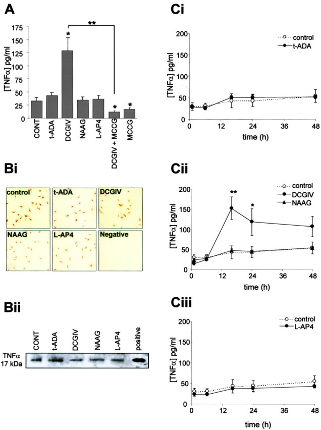
Activation of mGlu2 induces TNFα release from microglia. A, Microglial cells were exposed to the specific mGlu receptor agonist t-ADA (group I; 250 μm), DCGIV [group II; 500 nm; with or without group II antagonist MCCG (500 μm)], NAAG (mGlu3; 50 μm), l-AP-4 (group III; 100 μm), or MCCG (500 μm). After 24 h, the TNFα concentration in the culture medium was measured by ELISA. Data are the mean ± SEM from four to five separate experiments (defined as different cell preparations) per condition in which the replicates per experiment were three to four (defined as the number of separate coverslips for each condition). Bi, Immunolocalization of TNFα expression in control microglia and microglia exposed to mGlu receptor agonists, as above, for 24 h. Fixed cells were stained with a specific antibody to TNFα (1:500). Negative controls were treated in the same way, but the primary antibody was omitted. Bii, Cell lysates (45 μg of protein) from microglia exposed to mGlu receptor agonists for 24 h were subjected to immunoprecipitation for TNFα, and the immunocomplexes were resolved by 10% SDS-PAGE. Positive, Rat recombinant TNFα (1 μg/ml). C, Time course of TNFα release from microglia. Cells were exposed to the specific mGlu receptor agonists t-ADA (Ci; 250 μm), DCGIV (Cii; 500 nm), NAAG (Cii; 50 μm), and l-AP-4 (Ciii; 100 μm). At 1, 6, 16, 24, and 48 h after exposure, the culture medium was removed and the TNFα concentration was measured by ELISA. For each time point, values are mean ± SEM from four to five separate batches of cells with replicates of three to four separate coverslips. Significance values in all graphs are compared with control untreated MGCM (*p < 0.05; **p < 0.005), unless indicated otherwise.