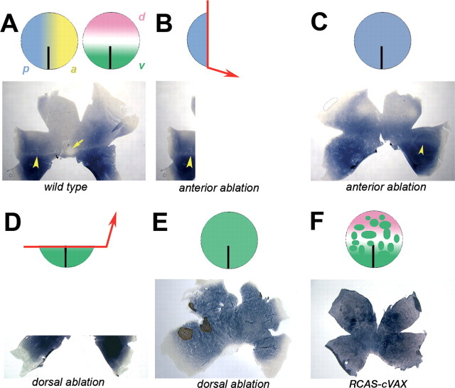
Figure 7.
Altered rod distribution after resection of the anterior eye anlage. A, The optic vesicle, schematically represented as circles, is polarized along the AP of the vesicle by several known and unknown factors. Anterior positional values are indicated by a yellow gradient, and posterior values are indicated by a blue gradient. Along the DV axis, the eye anlage is separated into the domains of expression of cVax ventrally (green) and Tbx5 dorsally (pink). Cells along the horizontal meridian of the optic vesicle express neither molecule. The normal rod pattern develops under the influence of these factors. B, Removal of the anterior eye anlage will leave a remnant of the optic vesicle behind of almost maximal posterior characteristics. These cells can only give rise to the posterior-most aspect of the rod pattern. C, When the optic vesicle reconstitutes by proliferation of the remaining cells, the cells appear to retain the positional information they had before the surgery. Consequently, the vesicle remnant gives rise to a retina in which the rod distribution resembles a repetition of the most posterior aspect of the pattern: a dorsal shift of the rod-rich ventral domain containing arod-sparse streak but no rod-free zone. D, Removal of the dorsal optic vesicle leaves a ventral remnant of the vesicle behind. E, After reconstitution, the healed optic vesicle is entirely composed of cells of ventral origin. Hence, the retina developing from such a vesicle is ventralized with respect to the rod distribution. F, Retroviral misexpression of cVax generates random patches of ectopic cVax expression scattered across the optic vesicle and thereby creates numerous new boundaries between cells expressing high levels of cVax and cells not expressing cVax. This leads to an altered distribution of rods across the retina.

