
Figure 1.
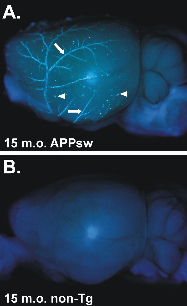
Ex vivo image of an APPsw brain demonstrating extensive CAA. A, A 15-month-old (15 m.o.) APPsw mouse brain stained with X-34 and imaged with UV epifluorescence to denote fibrillar amyloid. Extensive CAA in leptomeningeal vessels can be seen on the cortical surface (arrows) as well as on plaques near the cortical surface (arrowheads). B, A 15-month-old non-transgenic (non-Tg) mouse brain stained with X-34, demonstrating the specificity of the stain.