
Figure 2.
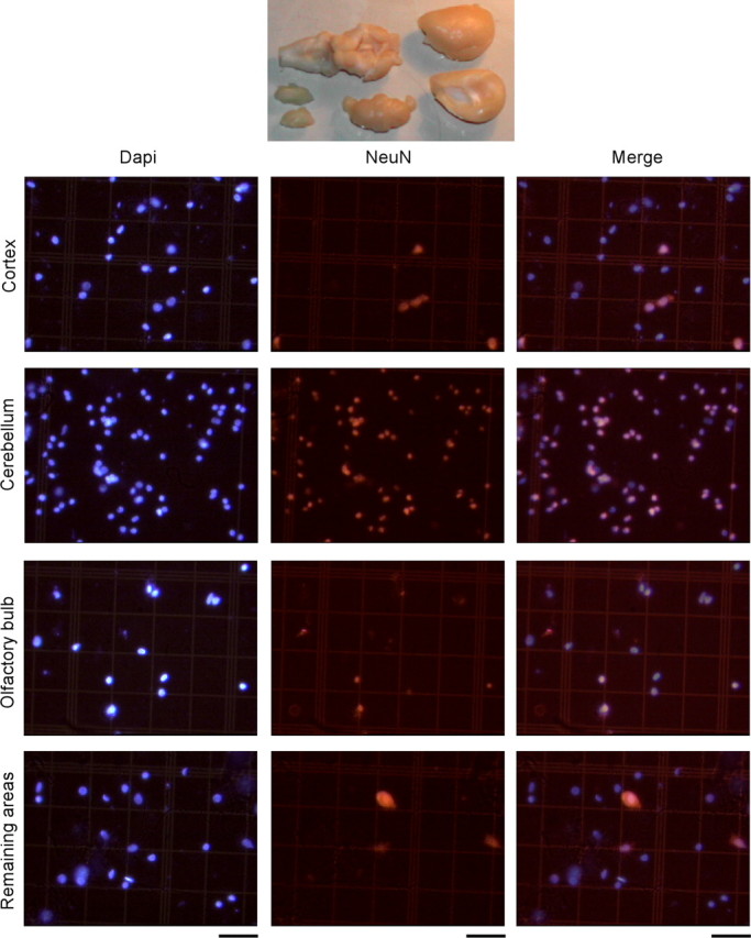
The total number of neurons in dissected brain regions (top; cortex, cerebellum, olfactory bulb, and remaining areas) is estimated by determining the proportion of DAPI-stained nuclei in isotropic suspensions (left) that are also NeuN positive (center; right, merged images). All of the photomicrographs are shown at the same magnification. Scale bars, 50 μm. Notice that the proportion of NeuN-positive nuclei is distinct among the four regions of interest. “Cortex” refers to gray and white matter of neocortex, hippocampus, and adjacent paleocortices lateral to the olfactory tract; “Cerebellum” refers to cerebellar cortex, deep nuclei, and cerebellar white matter, including the cerebellar peduncles. “Remaining areas” include accessory olfactory nuclei, basal ganglia, diencephalon and optic chiasm, and brainstem. Smaller, precise regions can also be quantified using the isotropic fractionator, provided constant criteria are used for dissection (e.g., specific cortical areas and gyri, subcortical nuclei, basal ganglia, diencephalon, midbrain, pons, and medulla). In addition, dissections can also be performed from vibratome sections.