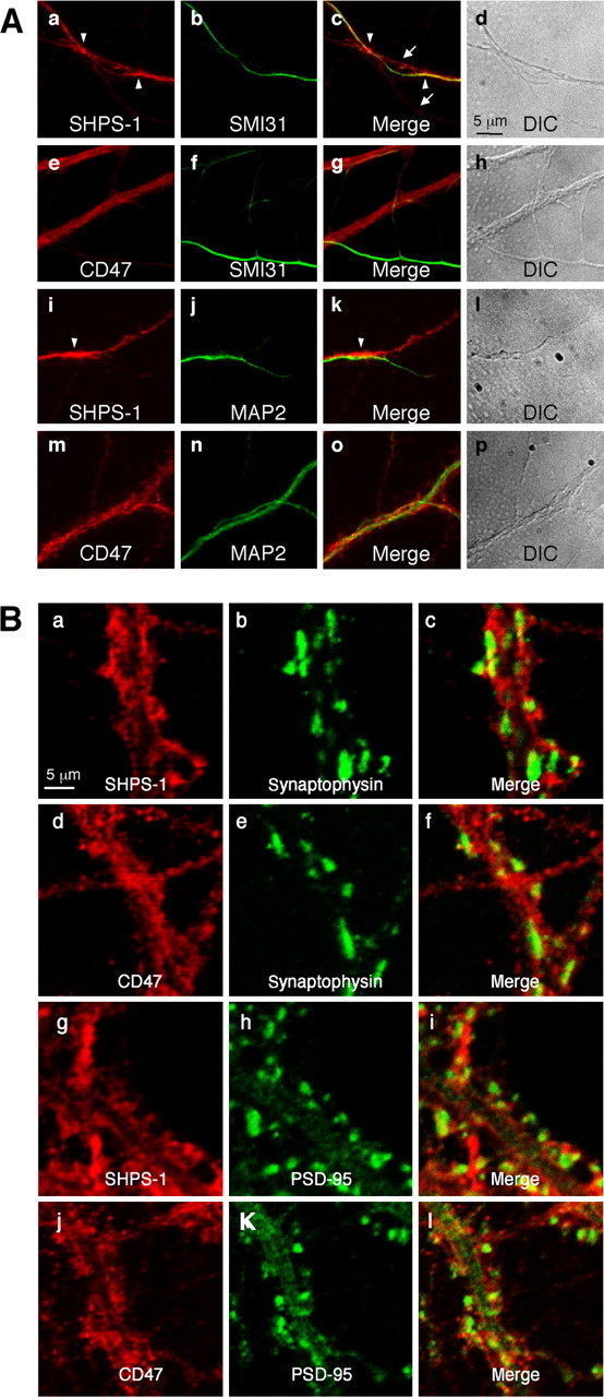
Figure 2.

Localization of endogenous SHPS-1 and CD47 in mouse hippocampal neurons at 20 or 28 DIV. A, Cultured mouse hippocampal neurons were fixed at 20 DIV and then subjected to two-color immunofluorescence staining with the indicated pairs of mAbs. c, g, k, and o are merged images of a and b, e and f, i and j, and m and n, respectively. Differential interference contrast (DIC) images corresponding to a-c, e-g, i-k, and m-o are shown in d, h, l, and p, respectively. The arrows in c indicate SHPS-1 immunoreactivity that was detected along SMI31-negative neurites. Arrowheads in a and c indicate intense SHPS-1 immunoreactivity at sites of contact between an SMI31-positive axon and the SMI31-negative dendrite-like neurite. The arrowheads in i and k indicate intense SHPS-1 immunoreactivity at sites of interaction between an SHPS-1-positive fiber and a MAP2-positive dendrite. B, Neurons were fixed at 28 DIV and then subjected to two-color immunofluorescence staining with the indicated pairs of mAbs. c, f, i, and l are merged images of a and b, d and e, g and h, and j and k, respectively. Scale bars, 5 μm. All results are representative of three separate experiments.
