Figure 3.

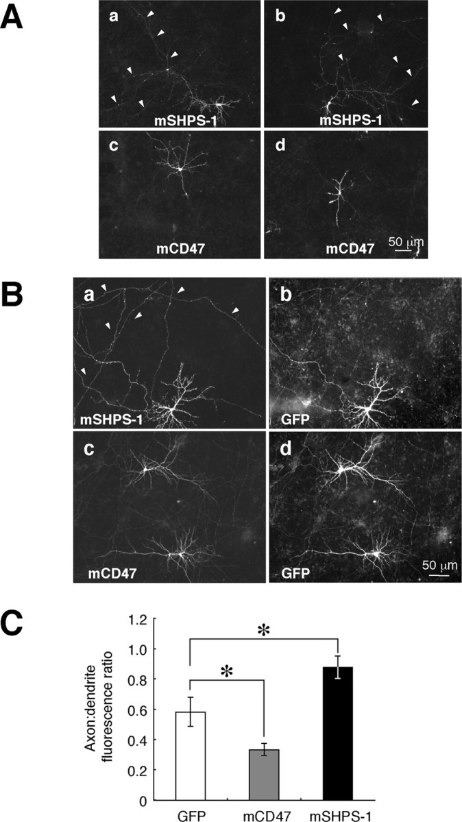
Localization of exogenously expressed mouse SHPS-1 and mouse CD47 in cultured rat hippocampal neurons. A, Neurons were transfected with expression vectors for mouse SHPS-1 (mSHPS-1) or mouse CD47 (mCD47), as indicated, at 12 DIV and fixed and immunostained with mAbs to mSHPS-1 or to mCD47, respectively, at 14 DIV. Arrowheads indicate long, thin fibers. B, Neurons were cotransfected with expression vectors for GFP and either mSHPS-1 (a, b) or mCD47 (c, d) at 10 DIV and fixed and stained with mAbs to mSHPS-1 (a) or to mCD47 (c) at 14 DIV. Fluorescence signals of GFP are shown in b and d. Arrowheads indicate long, thin fibers that were labeled intensely with the mAb to mSHPS-1. Scale bars, 50 μm. The results in A and B are representative of three separate experiments. C, Quantitation of the distribution of exogenously expressed GFP, mCD47, and mSHPS-1 in transfected neurons. The axon/dendrite fluorescence ratio was calculated for each protein as described in Materials and Methods. Data are means ± SE of values from 10 to 15 different cells. *p < 0.05 for the indicated comparisons (Student's t test).
