
Figure 4.
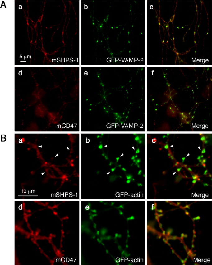
Localization of exogenously expressed SHPS-1 or CD47 relative to that of synaptic marker proteins in cultured rat hippocampal neurons. Neurons were cotransfected with expression vectors for GFP-VAMP-2 (A) or GFP-actin (B) and either mouse SHPS-1 (mSHPS-1) (a-c) or mCD47 (d-f) at 14 DIV. Seven days after transfection, the neurons were fixed and stained with mAbs to mSHPS-1 (a) or to mCD47 (d). The fluorescence signals of GFP-VAMP-2 (A) or GFP-actin (B) are shown in b and e. Merged images are shown in c and f. Arrowheads in Ba-c indicate intense CD47 immunoreactivity at dendritic spine sites marked with GFP-actin. Scale bars: A, 5 μm; B, 10 μm. All results are representative of three separate experiments.