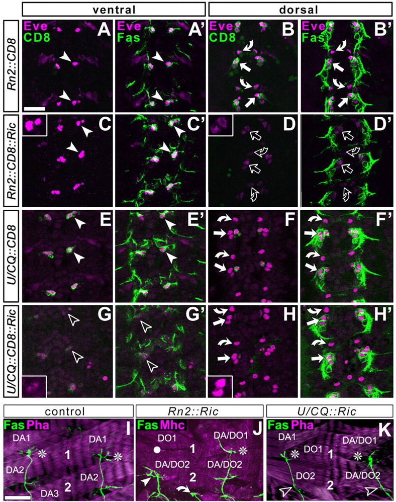
Figure 3.

Ablation studies using targeted expression of ricinA. The CNS at embryonic stage 13 (A-H′) or dorsal muscle fields at late stage 17 (I-K) are labeled as indicated at the top left: CD8, Gal4-induced mCD8-GFP (green in first and third column); Eve, the transcription factor Even-skipped (magenta in A-H′); Fas, FasciclinII (green in second and fourth column and bottom row); Mhc, the muscle marker myosin heavy chain (magenta in J); Pha, actin labeled with phalloidin (magenta in I, K). A-H′, Views of the horizontal plane of an abdominal CNS (anterior up) at embryonic stage 13 (the early motor axonal growth phase) visualized at ventral (left two columns) or dorsal (right two columns) levels, respectively, as indicated in the boxes at the top. Cell bodies of ventral U neurons (arrowheads), dorsal aCCs (straight arrows), and dorsal RP2s (curved arrows) were visualized with three independent markers: FasciclinII and Even-skipped are expressed in all of these neurons (Grenningloh et al., 1991; Broadus et al., 1995); mCD8-GFP is expressed only in those neurons targeted by the respective Gal4 line (genetic constellation indicated on left side). RN2-Gal4 targets CD8 expression to dorsal aCC/pCC and RP2 neurons (B) but not to ventral U neurons (A); U/CQ-Gal4 mediated expression occurs in ventral U neurons (E) but not in aCC/pCC and RP2 neurons (F). If these Gal4 lines are used to coexpress ricinA (Ric) with CD8, expression of all three markers is severely affected, but only in the targeted neurons (open symbols inD, D′ and G, G′; see especially the granular and weak occurrence of Eve; insets); furthermore, the Fasciclin II-labeled motor nerve is much thinner, especially after ablation of the larger group of U neurons (data not shown). I-K, Dorsal muscle fields of two consecutive hemisegments, respectively, at the end of embryogenesis in control animals or specimens with neuron-specific expression of ricinA. 1 and 2 indicate motor neuronal terminals on DA1/DO1 and DA2/DO2 muscles, respectively (some muscles indicated for orientation; compare Fig. 1). In controls and many cases of ricinA-ablated neurons, the ISN (shown with FasciclinII, green) grows to full length (white asterisks); in 26% of cases in which a CC/RP2 are ablated, ISNs stall in dorsolateral areas (white arrowhead in J; white circles indicate noninnervated muscles), whereas only 2.9% of ISNs stall in the case of U ablation (K, open arrowheads indicate absence of U terminals on DA/DO2 muscles); curved arrow indicates transverse nerve in J. Scale bar: (in A) A-H′, 20 μm; (in I) I-K, 16 μm.
