
Figure 3.
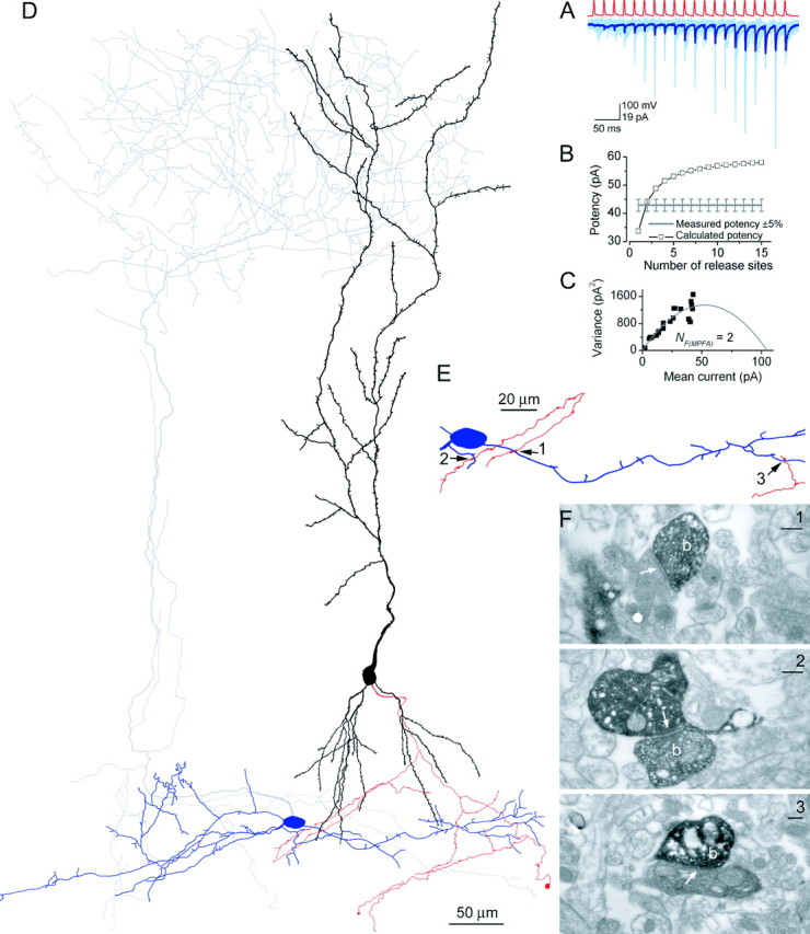
Functional and structural determination of the number of release sites for a PC-IN pair. A, Trains of presynaptic APs (red traces) evoked EPSCs in the postsynaptic IN (pair, AB377), which showed robust short-term facilitation (individual traces, light blue; averaged trace, dark blue). B, Potency amplitudes were calculated with a simple binomial model for integer Ns from 1 to 15. The calculated potency at N = 2 was almost identical to the measured potency at the end of the stimulus train, yielding NF(f) of 2. C, Multiple probability fluctuation analysis with a multinomial model also resulted in an NF(MPFA) of 2. The quantal size is 39.6 pA, and the Pr ranges from 0.07 to 0.39. D, LM reconstruction of the biocytin-labeled cells. The soma and dendrites of the presynaptic PC are shown in black; its axon arbor is red. The IN is classified as an O-LM cell based on the location of the soma and dendrites (blue) in the alveus and the extensive arborizations of its axon (gray) in the stratum lacunosum-moleculare. E, Locations of the three synaptic contacts (arrows) are shown at a higher magnification. Only parts of the IN and the PC are shown that establish the contacts. F, Electron microscopic images of the synaptic junctions (arrows) between three PC axon terminals (b) and the postsynaptic dendrites. Scale bars, 0.2 μm.