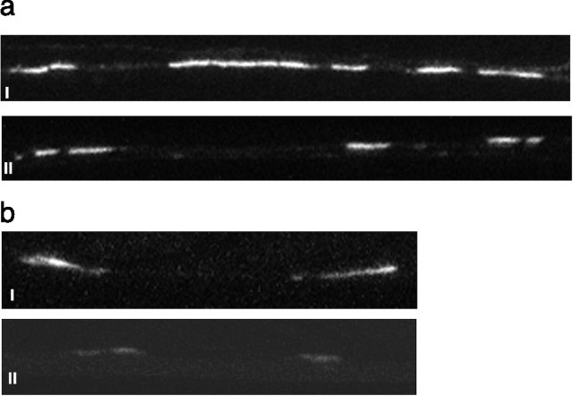
Figure 4.
Changes in glutamate receptor and vesicle concentration. a, Effects of isolation on GLR-1::GFP expression in the ventral nerve cord. Representative confocal image of a segment of the ventral nerve of a colony-raised worm (i) and an isolate-raised worm (ii) expressing GLR-1::GFP. White areas are clusters of GLR-1::GFP expressed in the ventral nerve cord. These are probably on or in the processes of interneurons of the tap withdrawal response (26). Isolate-raised worms showed less total GLR-1::GFP expression per image than did colony-raised worms. b, Effects of isolation on pmec-7::SNB-1::GFP expression in the ventral nerve cord. A representative confocal image of a segment of the ventral nerve cord of a colony-raised worm (i) and an isolate-raised worm (ii) expressing pmec-7::SNB-1::GFP is shown. White areas are GFP expressed in the sensory terminals of the tail sensory neurons (PLML and PLMR; 27). Isolate-raised worms showed significantly less pmec-7::SNB-1::GFP expression in the ventral cord than did colony-raised worms.

