
Figure 5.
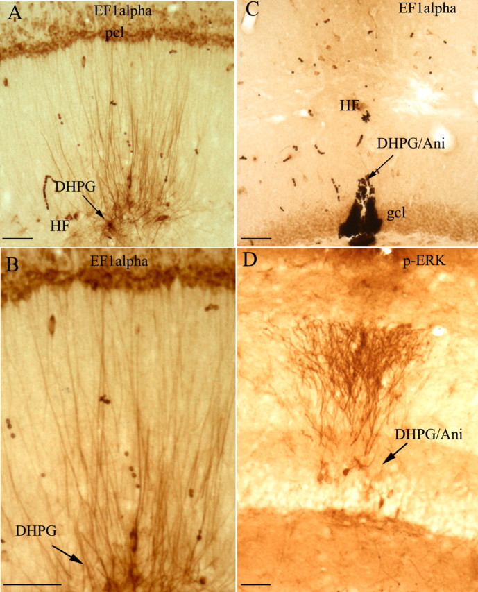
Local application of DHPG triggers a dramatic increase in EF1α protein levels in dendrites. A, The photomicrograph illustrates the striking increases in immunostaining for EF1α protein in dendrites surrounding a DHPG-filled micropipette (30 min after placement of the micropipette). B, High-power view of EF1α protein increase in the dendrites, which shows increased staining of dendrites with minimal if any increases in immunostaining at the level of the cell body, documenting that the increases in EF1α protein occur locally in dendrites. C, Increases in immunostaining for EF1α are blocked by systemic injection of the protein synthesis of anisomycin. Note the complete lack of any increase in dendritic staining in the area surrounding the micropipette. D, As a positive control for the efficacy of DHPG, a nearby section was immunostained for p-ERK. Note striking increases in p-ERK staining in the area surrounding the DHPG-filled micropipette in the DHPG/anisomycin experiment. Arrows indicate the path of the DHPG-filled micropipette. Scale bars, 50 μm. DHPG/Ani, DHPG/anisomycin; gcl, granule cell layer; HF, hippocampal fissure; pcl, pyramidal cell layer.