
Figure 7.
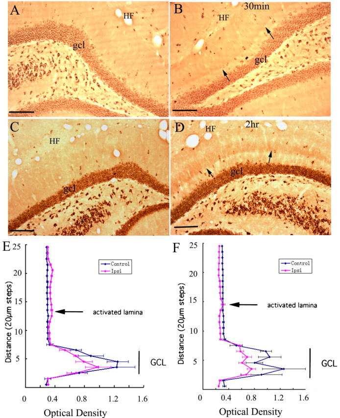
High-frequency stimulation of the medial perforant pathway alters immunostaining patterns for EF1α in the activated dentate gyrus. A, Pattern of immunostaining in the dentate gyrus on the side contralateral to the stimulation. B, Pattern of immunostaining after 30 min of high-frequency stimulation of the perforant path; small arrows indicate the band of increased immunostaining in the middle molecular layer (the site of termination of the medial perforant path). C, Pattern of immunostaining for EF1α on the control side of an animal that received 2 h of high-frequency stimulation. D, Pattern of immunostaining after 2 h of high-frequency stimulation. The graphs in E plot the average OD of EF1α immunostaining across the molecular layer from the cases illustrated in A and B, after 30 min of high-frequency stimulation, and F is the case illustrated in C and D, after 2 h of high-frequency stimulation. Error bars indicate the SD of the five measurements at each level. HF, Hippocampal fissure; gcl, granule cell layer. Arrows point to the activated dendritic lamina. Scale bars, 100 μm.