
Figure 6.
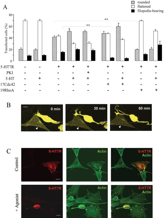
Regulation of cell morphology by the 5-HT7 receptor in NIH3T3 cells. NIH3T3 cells were transiently transfected with either a control (pTracer) vector or with pTracer vector encoding for the 5-HT7 receptor. Cells were cultured in serum-free medium overnight, and morphology was assessed after stimulation with agonists as indicated. A, Effect of the 5-HT7 receptor expression (with or without agonist stimulation) on the morphology of NIH3T3 cells. Dominant-negative mutants RhoA(N19) and Cdc42(N17) were coexpressed with the 5-HT7 receptor, as indicated. In several experiments, cells were treated with 10 μm cell-permeable PKA inhibitor 14-22 amide (PKI). Data points represent the means ± SEM from at least four independent experiments performed in duplicate. A statistically significant difference between values is indicated (*p < 0.05; **p < 0.01). B, Changes in the shape of cells expressing 5-HT7 receptors. NIH3T3 cells were transfected with pTracer vector encoding for the 5-HT7 receptor. Images shown were recorded by the confocal fluorescence microscopy at the beginning (0 min), during (30 min), and at the end (60 min) of a 1 h exposure to the 1 μm 5-HT with LSM510-Meta microscope at 63× magnification. A nontransfected cell is indicated by a triangle. Scale bar, 10 μm. The video is available as supplemental material (available at www.jneurosci.org). C, Analysis of the actin cytoskeleton in 5-HT7 receptor-expressing cells. NIH3T3 cells were transfected with GFP-tagged 5-HT7 receptor, serum starved, and then subjected to FITC-phalloidin staining. Representative confocal images obtained with LSM510-Meta microscope at 63× magnification are shown. Scale bar, 10 μm.