
Figure 7.
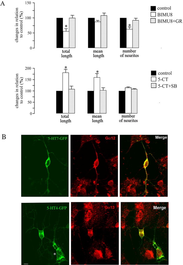
Effect of the 5-HT7R/G12 and 5-HT4R/G13 signaling pathways on neuronal morphology. A, For morphometric analysis of neurite outgrowth, cultured hippocampal neurons were fixed, stained with toluidine blue, and used for operator-controlled tracing of neurites. BIMU8 used at 100 nm is a selective 5-HT4 receptor agonist. GR113808 (GR) used at 2 μm is a highly selective 5-HT4 receptor antagonist. 5-CT used at 100 nm is a selective 5-HT7 receptor agonist. Treatment of the cells with the 5-HT7 receptor antagonist SB269970 (SB) at 100 nm blocked the effect of 5-CT. Data sets were collected after 20 h of treatment with an agonist/antagonist. The bars represent means ± SEM from three independent experiments performed in triplicate. Values obtained for untreated neurons were set to 100%. A statistically significant difference between values is indicated (*p < 0.05). B, Distribution of 5-HT7 and 5-HT4 receptors and Gα12 and Gα13 proteins in hippocampal neurons. The neurons were transfected with either GFP-tagged 5-HT4 or 5-HT7 receptors. The endogenous Gα12 or Gα13 proteins were visualized by immunostaining with specific antibodies. The asterisk indicates a transfected glial cell. Scale bar, 10 μm.